- Healthcare
Effectiveness of convalescent plasma therapy in severe COVID-19 patients

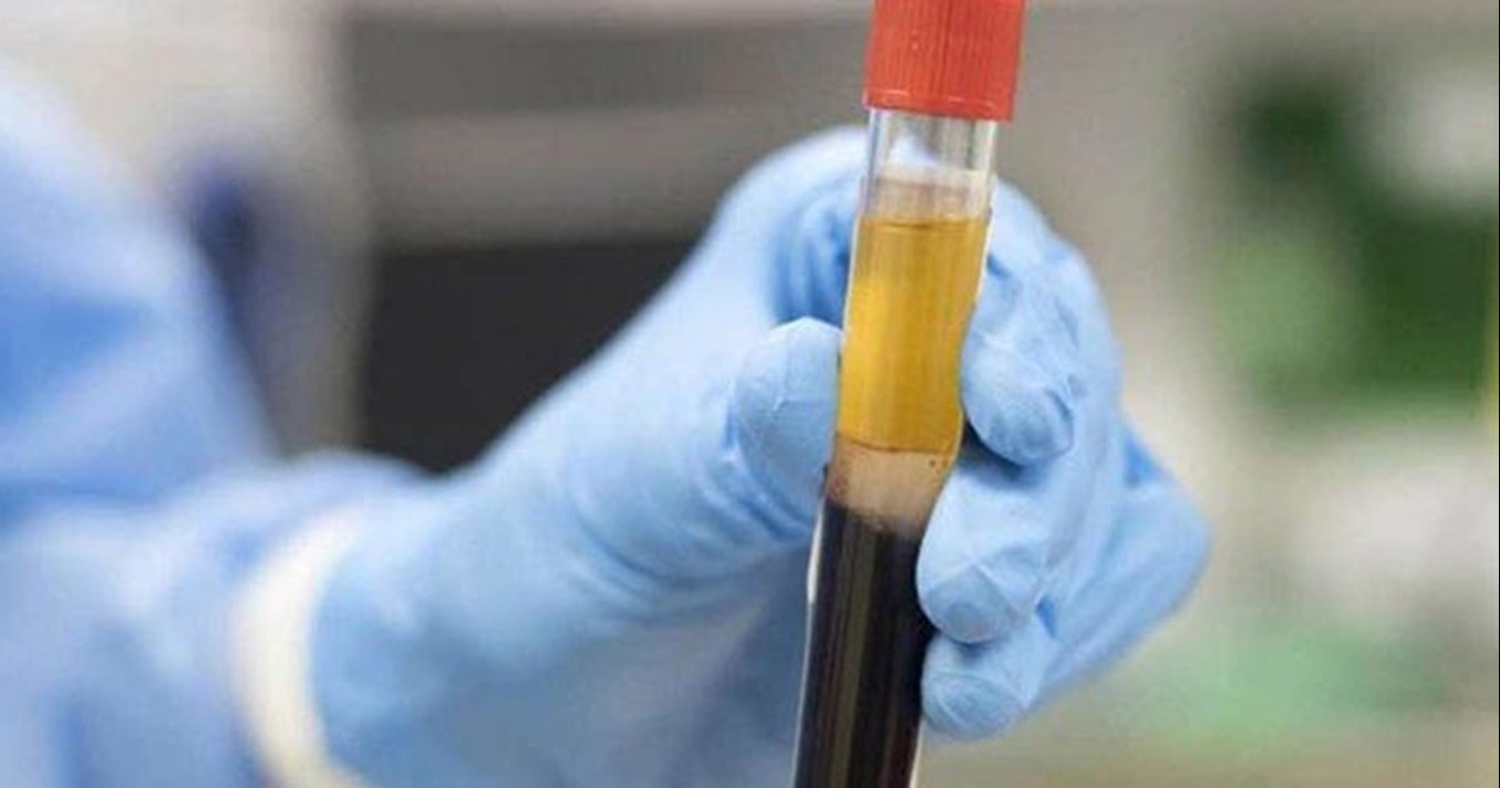
Convalescent plasma (CP) therapy has been administered as a classic adaptive immunotherapy for the prevention and treatment of many infectious diseases for over a century.
Duan et al., PNAS, 2020
Over the past two decades, CP therapy was successfully used in the treatment of SARS, MERS, and the 2009 H1N1 pandemic with satisfactory efficacy and safety1–4. A meta-analysis from 32 studies on SARS coronavirus infection and severe influenza showed a statistically significant reduction in the pooled odds of mortality following CP therapy, compared with placebo or no therapy (odds ratio, 0.25; 95% confidence interval, 0.14–0.45)5. However, the CP therapy proved to be ineffective at significantly improving the survival in the Ebola virus disease. The failure has been deemed to be due to the absence of data on the neutralizing antibody titration for stratified analysis6. Since the virological and clinical characteristics share similarity among SARS, Middle East Respiratory Syandrome (MERS), and COVID-197, it is clinically argued that CP therapy might be a treatment option for COVID-19 rescue8. Patients who have recovered from COVID-19 with a high neutralizing antibody titre may be a valuable donor source of CP. Nevertheless, the potential clinical benefit and risk associated with convalescent blood products in COVID-19 remain unproven. Duan and colleagues have now performed a pilot study in three participating hospitals to explore the feasibility and efficacy of CP treatment in 10 patients suffering from severe form of COVID-19.
In this study, the 10 severe patients confirmed by RT viral RNA test (RT-PCR) were enrolled prospectively. The most common symptoms at disease onset were fever (7 of 10 patients), cough (eight cases), and shortness of breath (eight cases), while less common symptoms included sputum production (five cases), chest pain (two cases), diarrhea (two cases), nausea and vomiting (two cases), headache (one case), and sore throat (one case). Four patients had underlying chronic diseases, including cardiovascular and/or cerebrovascular diseases and essential hypertension. Nine patients received arbidol monotherapy or combination therapy with remdesivir (in one case not included in the current clinical trial), or ribavirin, or peramivir, while one patient received ribavirin monotherapy. Antibacterial or antifungal treatment was used when patients had coinfection. Six patients received intravenous methylprednisolone (20 mg every 24 h). On computer-assisted tomography (CT), all patients presented bilateral ground-glass opacity and/or pulmonary parenchymal consolidation with predominantly subpleural and bronchovascular bundles distribution in the lungs. Seven patients had multiple lobe involvement, and four patients had interlobular septal thickening.
One dose of 200 mL of CP obtained and processed from recently recovered donors with the neutralizing antibody titres above 1:640 was transfused to the patients as an addition to maximal supportive care and antiviral agents. The primary endpoint was the safety of CP transfusion. The second endpoints were the improvement of clinical symptoms and laboratory parameters within 3 days post CP transfusion. The median time from onset of illness to CP transfusion was 16.5 days. After CP transfusion, the authors observed that the level of neutralizing antibody increased rapidly up to 1:640 in five cases, while that of the other four cases maintained at a high level (1:640). The clinical symptoms markedly improved along with increase of oxyhemoglobin saturation within 3 days. It is noteworthy that several clinical parameters tended to improve as compared to pretransfusion, including increased lymphocyte counts (0.65 × 10⁹ /L vs. 0.76 × 10⁹ /L) and decreased C-reactive protein (55.98 mg/L vs. 18.13 mg/L). Radiological examinations showed varying degrees of absorption of lung lesions within 7 days. The viral load was undetectable after transfusion in seven patients who had previous viremia. No severe adverse effects were observed.
Severe pneumonia caused by human coronavirus was characterized by rapid viral replication, massive inflammatory cell infiltration, and elevated proinflammatory cytokines or even cytokine storm in alveoli of lungs, resulting in acute pulmonary injury and acute respiratory distress syndrome (ARDS)9. Recent studies on COVID-19 demonstrated that the lymphocyte counts in the peripheral blood were remarkably decreased and the levels of cytokines in the plasma from patients requiring intensive care unit (ICU) support, including IL-6, IL-10, TNF-ɑ, and granulocyte-macrophage colony-stimulating factor, were significantly higher than in those who did not require ICU conditions10. CP, obtained from recovered COVID-19 patients who had established humoral immunity against the virus, contained a large quantity of neutralizing antibodies capable of neutralizing SARS-CoV-2 and eradicating the pathogen from blood circulation and pulmonary tissues11. In the present study, all investigated patients achieved serum SARS-CoV-2 RNA negativity after CP transfusion, accompanied by an increase of oxygen saturation and lymphocyte counts, and the improvement of liver function and CRP. The results suggest that the inflammation and overreaction of the immune system were alleviated by antibodies contained in CP. The case fatality rates (CFRs) in the present study were 0% (0/10), which was comparable to the CFRs in SARS, which varied from 0% (0/10) to 12.5% (10/80) in four noncomparative studies using CP treatment12-14. Based on our preliminary results, CP therapy can be an easily accessible, promising, and safe rescue option for severe COVID-19 patients. It is, nevertheless, worth mentioning that the absorption of pulmonary lesions often lagged behind the improvement of clinical symptoms, as shown in patients 9 and 10 in this trial.
The first key factor associated with CP therapy is the neutralizing antibody titre. A small sample study in MERS-CoV infection showed that the neutralizing antibody titre should exceed 1:80 to achieve effective CP therapy4. To find eligible donors who have high levels of neutralizing antibody is a prerequisite. Cao et al.15 showed that the level of specific neutralizing antibody to SARS-CoV decreased gradually 4 months after the disease process, reaching undetectable levels in 25.6% (IgG) and 16.1% (neutralizing antibodies) of patients at 36 months after disease status. A study from the MERS-CoV−infected patients and the exposed healthcare workers showed that the prevalence of MERS-CoV IgG seroreactivity was very low (2.7%), and the antibodies titre decreased rapidly within 3 months16. These studies suggest that the neutralizing antibodies represent short-lasting humoral immune response, and plasma from recently recovered patients should be more effective. In the present study, recently recovered COVID-19 patients, who were infected by SARS-CoV-2 with neutralizing antibody titre above 1:640 and recruited from local hospitals, were considered as suitable donors. The median age of donors was lower than that of recipients (42.0 y vs. 52.5 y). Among the nine cases investigated, the neutralizing antibody titres of five patients increased to 1:640 within 2 days, while four patients kept the same level. The antibody titres of CP in COVID-19 seem higher than those used in the treatment of MERS patient (1:80)4.
The second key factor associated with efficacy is the treatment time point. A better treatment outcome was observed among SARS patients who were given CP before 14 days (58.3% vs. 15.6%; P < 0.01), highlighting the importance of timely rescue therapy (9). The mean time from onset of illness to CP transfusion was 16.5 days. Consistent with previous research, all three patients receiving plasma transfusion given before 14 days (patients 1, 2, and 9) in the study showed a rapid increase of lymphocyte counts and a decrease of CRP, with remarkable absorption of lung lesions in CT. Notably, patients who received CP transfusion after 14 days showed much less significant improvement, such as patient 10. However, the dynamics of the viremia of SARS-CoV-2 was unclear, so the optimal transfusion time point needs to be determined in the future. In the present study, no severe adverse effects were observed. One of the risks of plasma transfusion is the transmission of the potential pathogen. Methylene blue photochemistry was applied in this study to inactivate the potential residual virus and to maintain the activity of neutralizing antibodies as much as possible, a method known to be much better than ultraviolet (UV) C light17. No specific virus was detected before transfusion. Transfusion-related acute lung injury was reported in an Ebola virus disease woman who received CP therapy18.
Although uncommon in the general population receiving plasma transfusion, the specific acute lung injury adverse reaction is worth noting, especially among critically ill patients experiencing significant pulmonary injury19. Another rare risk worth mentioning during CP therapy is antibody-dependent infection enhancement, occurring at subneutralizing concentrations, which could suppress innate antiviral systems and thus could allow logarithmic intracellular growth of the virus20. The special infection enhancement also could be found in SARS-CoV infection in vitro21. No such pulmonary injury and infection enhancement were observed in this study, probably because of the high levels of neutralizing antibodies, timely transfusion, and appropriate plasma volume. Overall, the study has a number of limitations. First, except for CP transfusion, the patients received other standard care. All patients received antiviral treatment despite the uncertainty of the efficacy of drugs used. As a result, the possibility that these antiviral agents could contribute to the recovery of patients, or synergize with the therapeutic effect of CP, could not be ruled out.
Furthermore, some patients received glucocorticoid therapy, which might interfere with immune response and delay virus clearance. Second, the median time from onset of symptoms to CP transfusion was 16.5 days. Although the kinetics of viremia during natural history remains unclear, the relationship between SARS-CoV-2 RNA reduction and CP therapy, as well as the optimal concentration of neutralizing antibodies and treatment schedule, should be further clarified. Third, the dynamic changes of cytokines during treatment were not investigated. Nevertheless, the preliminary results of this trial appear promising, justifying a randomized controlled clinical trial in a larger patient cohort. In conclusion, this pilot study on CP therapy shows a potential therapeutic effect and low risk in the treatment of severe COVID-19 patients. One dose of CP with a high concentration of neutralizing antibodies can rapidly reduce the viral load and improve clinical outcomes. The optimal dose and treatment time point, as well as the definite clinical benefits of CP therapy, need to be further investigated in randomized clinical studies.
This article is intended for medical professionals.
Read also:
An Evidence-based Perspective into Treatment options for COVID-19
Compassionate Use of Remdesivir for Patients with Severe COVID-19
References:
1. Y. Cheng et al., Use of convalescent plasma therapy in SARS patients in Hong Kong. Eur. J. Clin. Microbiol. Infect. Dis. 24, 44–46 (2005).
2. B. Zhou, N. Zhong, Y. Guan, Treatment with convalescent plasma for influenza A (H5N1) infection. N. Engl. J. Med. 357, 1450–1451 (2007).
3. I. F. Hung et al., Convalescent plasma treatment reduced mortality in patients with severe pandemic influenza A (H1N1) 2009 virus infection. Clin. Infect. Dis. 52, 447–456 (2011).
4. J. H. Ko et al., Challenges of convalescent plasma infusion therapy in Middle East respiratory coronavirus infection: A single centre experience. Antivir. Ther. 23, 617– 622 (2018).
5. J. Mair-Jenkins et al.; Convalescent Plasma Study Group, The effectiveness of convalescent plasma and hyperimmune immunoglobulin for the treatment of severe acute respiratory infections of viral etiology: A systematic review and exploratory metaanalysis. J. Infect. Dis. 211, 80–90 (2015).
6. J. van Griensven et al.; Ebola-Tx Consortium, Evaluation of convalescent plasma for Ebola virus disease in Guinea. N. Engl. J. Med. 374, 33–42 (2016).
7. P. I. Lee, P. R. Hsueh, Emerging threats from zoonotic coronaviruses-from SARS and MERS to 2019-nCoV. J. Microbiol. Immunol. Infect., in press.
8. L. Chen, J. Xiong, L. Bao, Y. Shi, Convalescent plasma as a potential therapy for COVID-19. Lancet Infect. Dis. 20, 398–400 (2020).
9. R. Channappanavar, S. Perlman, Pathogenic human coronavirus infections: Causes and consequences of cytokine storm and immunopathology. Semin. Immunopathol. 39, 529–539 (2017).
10. C. Huang et al., Clinical features of patients infected with 2019 novel coronavirus in Wuhan, China. Lancet 395, 497–506 (2020).
11. G. Marano et al., Convalescent plasma: New evidence for an old therapeutic tool? Blood Transfus. 14, 152–157 (2016).
12. V. W. Wong, D. Dai, A. K. Wu, J. J. Sung, Treatment of severe acute respiratory syndrome with convalescent plasma. Hong Kong Med. J. 9, 199–201 (2003).
13. K. M. Yeh et al., Experience of using convalescent plasma for severe acute respiratory syndrome among healthcare workers in a Taiwan hospital. J. Antimicrob. Chemother. 56, 919–922 (2005).
14. L. K. Kong, B. P. Zhou, Successful treatment of avian influenza with convalescent plasma. Hong Kong Med. J. 12, 489 (2006).
15. W. C. Cao, W. Liu, P. H. Zhang, F. Zhang, J. H. Richardus, Disappearance of antibodies to SARS-associated coronavirus after recovery. N. Engl. J. Med. 357, 1162– 1163 (2007).
16. Y. M. Arabi et al., Feasibility of using convalescent plasma immunotherapy for MERSCoV infection, Saudi Arabia. Emerg. Infect. Dis. 22, 1554–1561 (2016).
17. M. Eickmann et al., Inactivation of Ebola virus and Middle East respiratory syndrome coronavirus in platelet concentrates and plasma by ultraviolet C light and methylene blue plus visible light, respectively. Transfusion 58, 2202–2207 (2018).
18. M. Mora-Rillo et al.; La Paz-Carlos III University Hospital Isolation Unit, Acute respiratory distress syndrome after convalescent plasma use: Treatment of a patient with Ebola virus disease contracted in Madrid, Spain. Lancet Respir. Med. 3, 554–562 (2015).
19. A. B. Benson, M. Moss, C. C. Silliman, Transfusion-related acute lung injury (TRALI): A clinical review with emphasis on the critically ill. Br. J. Haematol. 147, 431–443 (2009).
20. S. B. Halstead, Dengue antibody-dependent enhancement: Knowns and unknowns. Microbiol. Spectr. 2, AID-0022-2014 (2014).
21. S. F. Wang et al., Antibody-dependent SARS coronavirus infection is mediated by antibodies against spike proteins. Biochem. Biophys. Res. Commun. 451, 208–214 (2014).
Prepared by:
Dr Reshma Ramracheya
Research Scientist & University Research Lecturer at the University of Oxford
Senior Research Fellow at Wolfson College, University of Oxford
Reshma.ramracheya@ocdem.ox.ac.uk


